Cranial ultrasound a guideline for the performance of routine cranial USS for preterm infants
Ultrasound imaging of the head uses sound waves to produce pictures of the brain and cerebrospinal fluid. It is usually performed on infants, whose skulls have not completely formed. A transcranial Doppler ultrasound evaluates blood flow in the brain's major arteries. Ultrasound is safe, noninvasive, and does not use ionizing radiation.
Cranial Ultrasound UAMS Department of Radiology
Antiamyloid antibodies have been used to reduce cerebral amyloid-beta (Aβ) load in patients with Alzheimer's disease. We applied focused ultrasound with each of six monthly aducanumab infusions to temporarily open the blood-brain barrier with the goal of enhancing amyloid removal in selected brain regions in three participants over a period of 6 months.

Fetal Brain Anatomy
In the past three decades, cerebral ultrasound (CUS) has become a trusted technique to study the neonatal brain. It is a relatively cheap, non-invasive, bedside neuroimaging method available in.

Future applications of advanced neonatal cerebral ultrasound Paediatrics and Child Health
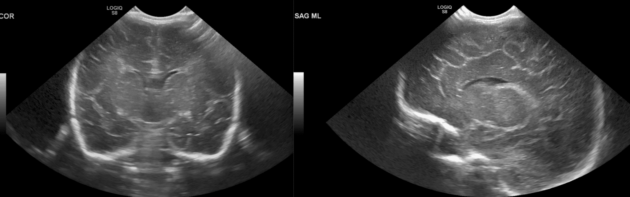
CrUS should be performed by a person who is familiar with the brain anatomy, brain maturation and commonly occurring abnormalities. Images should be labelled with patient identification, examination date, and image orientation. CrUS is performed with basic grey scale imaging. Understanding of ultrasound principles is required to interpret the.

Normal neonatal brain sonography of a preterm newborn in right lateral... Download Scientific
Although MRI is the reference standard for infant brain imaging, it is expensive, often requires sedation, and may not be possible to perform on critically ill patients.. North K, Lowe L. Modern head ultrasound: normal anatomy, variants, and pitfalls that may simulate disease. Ultrasound Clin 2009; 4:497 -512. Crossref. Google Scholar. 6.

Practical guide to neonatal cranial ultrasound (CrUS) basics Paediatrics and Child Health
Citation, DOI, disclosures and article data. Transcranial Doppler ( TCD ) , also known as transcranial color-coded duplex sonography ( TCCS) is a sonographic study of intracranial structures and blood vessels, used most commonly to identify the hemodynamic state present in the vertebrobasilar circulation and the circle of Willis .

Practical guide to neonatal cranial ultrasound (CrUS) basics Paediatrics and Child Health
Brain ultrasonography can be used to evaluate cerebral anatomy and pathology, as well as cerebral circulation through analysis of blood flow velocities.

Image
Our study explores the topography of the brain on B‐mode ultrasound in adult critically ill patients and provides descriptive examples of the anatomical and pathological structures that can be visualized on B‐mode cranial ultrasonography.

Practical guide to neonatal cranial ultrasound (CrUS) basics Paediatrics and Child Health
Several studies have shown that focused ultrasound can safely and transiently open the blood-brain barrier in patients with Alzheimer's disease and other neurologic disorders. 3-13 The effect.

Coronal section of brain The BMJ
Chronic abnormality of intracranial anatomy. intracranial hematoma (back to contents) Intracranial hematomas will appear hyperechoic for about five days. Subsequently, they will become hypoechoic, with a surrounding hyperechoic halo.. Venkatraghavan L. Clinical applications of point-of-care ultrasound in brain injury: a narrative review.

Normal neonatal brain sonography of a preterm newborn in sagittal... Download Scientific Diagram
Brain Anatomy on Cranial Ultrasound. B mode anatomy can visualize the midbrain in the axial plane visible as a "butterfly shape" with cerebral peduncles and colliculi. Figure 2a illustrates brain anatomy on ultrasound in a patient without a skull flap after undergoing hemicraniectomy for intracranial hemorrhage resection. Most patients with.

Practical guide to neonatal cranial ultrasound (CrUS) basics Paediatrics and Child Health
Head ultrasound (HUS), also called cranial ultrasound (CUS), is obtained for the diagnosis and follow-up of premature and sick neonates. Advantages Head ultrasound has the advantages of: accessibility mobility, i.e. bedside scanning at the NICU and neonatal ward requiring no sedation

Ultrasound and MR Imaging of the Normal Fetal Brain Neuroimaging Clinics
In this chapter, we will discuss ultrasound anatomy of the neonatal brain, how to differentiate normal findings from (subtle) abnormalities, the optimal timing of ultrasound examinations, and the most frequently occurring brain lesions of the preterm infant. 3.1 Ultrasound Anatomy of the Neonatal Brain

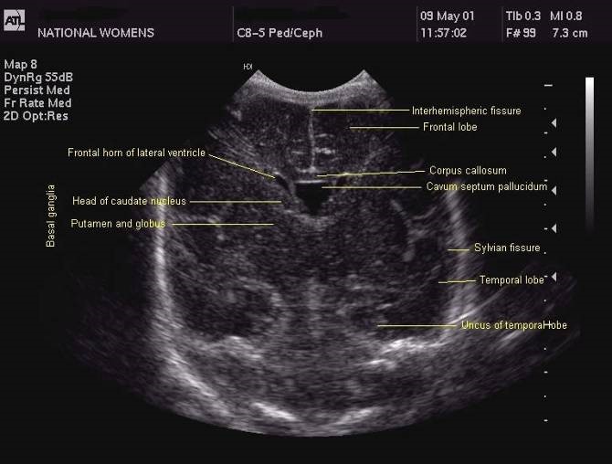
Infant Head Ultrasound Anatomy dbabyzi
Abstract Cranial ultrasound (CUS) is an extremely valuable tool to evaluate the brain during the first year of life, in experienced hands. It is the initial screening imaging tool to evaluate the infants' brain and complementary to the use of computed tomography (CT) and magnetic resonance imaging (MRI).

Ultrasound fetal brain image from left to right the columns show the... Download Scientific
00:37 Above. Normal fetal CNS at 22 2/7ths weeks. Video courtesy of Dr. Mayank Chowdhury; Pallav Imaging Institute, Mayflower Women's Hospital, Ahmedabad, India. Above. Key fetal anatomy includes the choroid plexus, the septum cavum pellucidi (SCP), the lateral ventricles, and the corpus callosum.
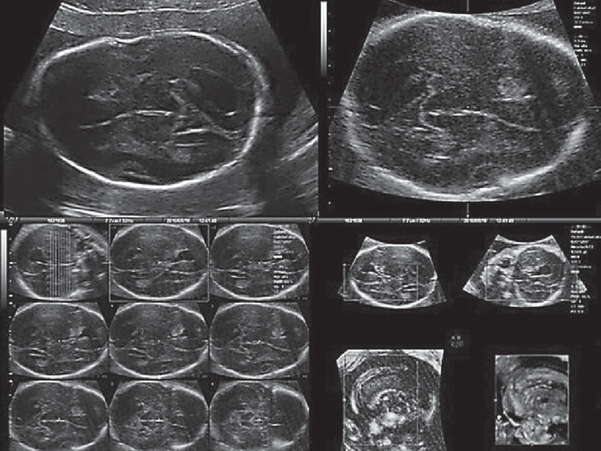
Case 2 2D and 3D fetal brain ultrasound showing marked ventricular... Download Scientific Diagram
Ultrasound is an exceptional tool for the evaluation of an infant's brain. It is portable and can be easily and quickly performed at the bedside. Therefore, the initial imaging modality used to evaluate the intracranial anatomy and identify intracranial pathologies such as congenital anomalies, intracranial hemorrhage, ischemia, and hydrocephalus, especially in sick preterm infants.